Turbo-BrainVoyager v4.4
Scan Parameters
The Scan tab (see screenshot below) of the Data Simulator is used to specify important parameters for generated slices, including the spatial and temporal (sampling) resolution of the simulated data.
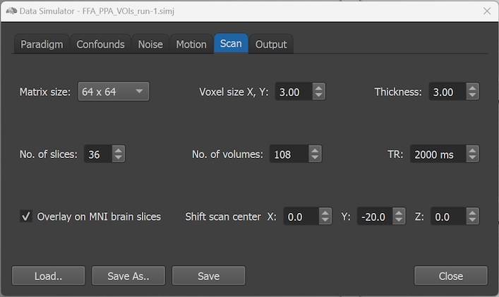
The Matrix size selection box can be used to select the dimensions of the generated images (slices). Clicking the selection box currently reveals 3 matrix sizes: 64 x 64 (selected as default), 96 x 96 and 128 x 128. The voxel size can be determined within plane using the Voxel size X, Y number field and along the third (slice) direction via the Thickness number field (there is no possibility to determine a gap between the slices). As default, iso-voxels are defined since both the inplane resolution and the slice thickness have the same default value (3.0). Note that the voxel size together with the matrix size determine the slice field-of-view (FOV), i.e. the extend of the brain covered along the slice dimensions. The coverage along the slice direction is determined by the number of scanned slices, which can be specified in the No. of slices number field (default: 36).
The generated number of functional volumes is determined by the No. of volumes number field. As soon as a protocol is selected in the Paradigm tab, it is used to set the value of the No. of volumes field automatically to the highest time point value (usually from the last 'baseline' condition that needs to be defined before the main experimental condition intervals). The temporal sampling resolution is determined by the volume-TR, which can be changed using the TR number field (default value: 2000 ms).
If checked (default), the option Overlay on MNI brain slices will slice a model brain in MNI space to generate EPI-like slices on which the functional data can be visualized when running, e.g., a standard (incremental) GLM. If the option is turned off, the regions will be placed at the same locations within the generated slices but no brain background will be visible. In addition to the EPI-like slices, the simulated data is also shown on a high-resolution T1-weighted brain when switching to the Anatomical Volume View in the main window of TBV. The Shift scan center X/Y/Z fields allow to move the scanned EPI slices relative to the center of MNI space. The values for the X and Z direction are set to 0.0 as default but the default value of the Y (posterior-to-anterior) axis is set to -20.0, which shifts the scanning along the Y axis in order to center the slices better in this direction.
Copyright © 2002 - 2024 Rainer Goebel. All rights reserved.