BrainVoyager v23.0
EEG and MEG File Formats
This section provides a general description of EEG/MEG data and auxiliary files that are needed for performing EEG/MEG distributed source modeling and analysis in BrainVoyager QX. Before starting the EMEG Suite, channel data and configurations and paradigm information are to be made available according to the BrainVoyager QX file format specifications.
Channel Data Sets
Channel time-course data are stored in CTC files and protocol information is stored in PRT files. CTC and PRT files are processed in the time and in the time-frequency domain resulting in three different new file types: averaged channel time-course data (ACT), single-trial data (STD) and time-frequency data (TFD) files. While CTCs contain continuous data, STDs and TFDs contain "epoched" data (although these are allowed to contain just one epoch). STDs are used for channel covariance calculations (Covariance Calculation) and the resulting covariance matrices are stored in CVX files.
There are two paths for importing new data sets in BrainVoyager QX:
1) Importing one single continuous channel time-courses in a CTC file with data from an entire EEG/MEG session or a segment. In this case a protocol PRT file is to be supplied containing all trigger information as conditions (condition = trigger type) and trials (with onset and offsets in microsecond time resolution). When using the EEG/MEG Data Import wizard, both CTCs and PRTs are normally generated automatically from the raw data files (see the EEG/MEG data import section for more details).
2) Importing single-trial data sets as one or more STD files corresponding to one or more different trigger types with epoched data from a given EEG/MEG session. This assumes that data have been already preprocessed and is normally achived with the help of some external tools.
EEG and MEG raw data sets from several file formats can be directly imported from the EEG-MEG menu (Import EEG/MEG Data ...) by invoking the EEG-MEG Data Import wizard. But, it is often possible to import EEG and MEG data sets from other EEG/MEG software packages with the help of simple functions and scripts running under Matlab 7 or later versions (www.mathworks.com) after installing a freely available toolbox called BVQXtools (http://support.brainvoyager.com/available-tools/52-matlab-tools-bvxqtools.html). Besides reading and writing all BrainVoyager QX file types, BVQXtoolsallows to create, read write all EEG/MEG BrainVoyager QX files. CTCs, PRTs and STDs can be created from data and trigger information files generated in BESA (see www.besa.de), EEGLAB (see http://sccn.ucsd.edu/eeglab/index.html), FIELDTRIP (see http://www.ru.nl/fcdonders/fieldtrip/fieldtrip) and NEUROMAG (see http://www.kolumbus.fi/kuutela/programs/meg-pd/). Further and more specific support about importing EEG and MEG data sets in BrainVoyager QX can be requested by contacting directly the BrainVoyager Support service (support@brainvoyager.com).
An important remark concerns the physical aspects of the signal scaling (units) and the configuration. Specifically, EEG sample values must be supplied in uV. If necessary, all EEG channel time-series must be average-referenced before entering the EMEG Suite (see the EEG/MEG data filter section). MEG sample values must be supplied in fT/cm according a 1-st order gradiometer configuration (see below).
Channel Configuration
Channel configuration data (CCD) is a file format used to record important information associated with EEG or MEG channels. CCD files are automatically generated at the time data are imported from external file formats. A CCD file contains linking information towards both a CTC and a PRT file, so that channel information is associated with the appropriate data and protocol. The CCD file header typically records general channel parameters before (i.e. original parameters) and after processing. These general parameters include data such as number of channels, sampling frequency, number of time samples and total duration of the data. Track is also kept for all supported preprocessing operations that were used on the dataset, such as selection of a channel subset, selection of a temporal subset, frequency filtering operations and rereferencing.
The body of the CCD file also contains specific information associated with each channel. This includes labels used to identify each channel, sensor geometry information used for computations relative to electromagnetic fields, digitization information used to convert between digital and physical units, sampling frequency, cut-frequencies for applied filters, and BrainVoyager specific display information.
A precise head/MRI coregistration is essential for distributed source modeling and analysis and can be performed in the surface module of BrainVoyager QX. This co-registration is achieved by importing fiducial points and head surface points in SFH files and invoking the BrainVoyager QX functions of the EEG-MEG menu. A surface reconstruction of the subject's head from a VMR covering the entire head is necessary for registration of the surface points. The result of this coregistration is an affine transformation whose parameters are appended to the same SFH (ASCII) file.
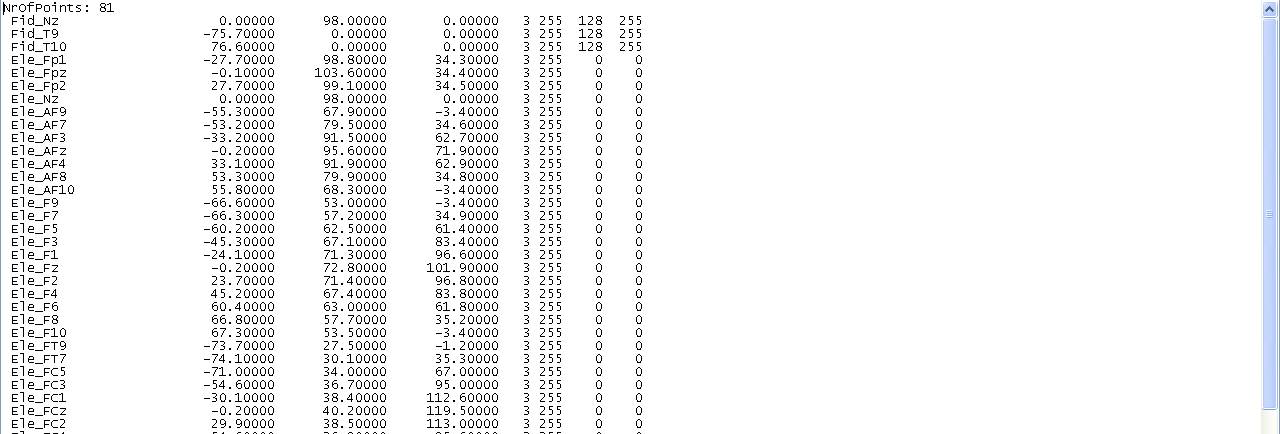
When analyzing EEG data, all electrodes are treated as surface points and vice versa (with the sole exclusion of the fiducial points). Therefore, the EEG channel coordinates are listed in the SFH file (after the three fiducials) and the EEG configuration is fully specified by the SFH file. An example of an SFH file used for specifying a 78-channel EEG configuration is shown below:


The first line reports the total number of surface points (3 fiducials + 78 electrodes). The three fiducials must be always in this order: Nasion, Left and Right Auricolar. Then the electrode come and X, Y and Z coordinates (in mm) are reported followed by other numbers controlling the display of the surface points. The coordinates are initially given i the head or device coordinate system. The final lines are automatically appended by the program after head/surface coregistration.
When analyzing MEG data, the SFH only contains surface points (e. g. positioning coils) useful for head-MRI egistration. In addition to the SFH, an ASCII-coded text file (with extension POS) is necessary and must contain the MEG primary and secondary sensor coordinates in [meter] physical units, together with their "shared" orientation. For MEG, a 1-st order gradiometer configuration is assumed and the POS file should only contain a series of lines, each line including 9 numbers, the X, Y and Z coordinates of the primary sensor, the X, Y and Z coordinates of the secondary sensor and the sensor (common) orientation.

Copyright © 2023 Fabrizio Esposito and Rainer Goebel. All rights reserved.